baby chest x ray radiation
Most X-ray exams including those of the legs head teeth or chest wont expose your reproductive organs to the direct X-ray beam and a lead apron can be worn to provide protection from radiation scatter. What are X-rays.

Chest X Ray Image Of A Patient With The Belt Fastened Around The Download Scientific Diagram
X-rays use a small amount of radiation about the same levels that occur naturally in the environment.
. The radiation exposure of an adjacent newborn the radiographer and other persons in the room was simulated using phantoms during X-ray examination of the chest using vertical and horizontal beams. Theres far less radiation exposure with an X-ray. A chest x-ray produces images of the heart lungs airways blood vessels and the bones of the spine and chest.
By comparison a chest x-ray delivers a. Diagnostic x-rays and other medical radiation procedures of the abdominal area also deserve extra attention during pregnancy. Chest X-rays are quick noninvasive tests.
Fetal doses resulting from radiological examination of the mothers skull head neck chest and extremities are extremely low 001 rad because of the relatively low maternal radiation dose beam direction and distance between the primary field and the fetus. A chest x-ray for example delivers 01 mSv while a chest CT delivers 7 mSv see the table 70 times as much. Erect chest X-rays are taken at 180 cm.
Risks depend on the amount of radiation to which the baby was exposed and the amount of time that it was exposed. Appointment Center 247 2164457050. For medical X-ray imaging the pediatric patients size is even more important to consider than age because patient size determines how much radiation is needed to.
X-rays are used to make pictures of the bones and organs. Free Healthy Baby App for iPhone. This amount of radiation is less than the.
One single chest x-ray is not concerning at all--even to newborns. A chest X-ray is a painless noninvasive procedure with few risks. X-rays are a form of radiation.
But the risk is very small. Your bones appear white because they are very dense. An x-ray exam helps doctors diagnose and treat medical conditions.
The part of the mothers body exposed to the radiation. Healthcare providers use chest X-rays to diagnose or treat conditions like pneumonia emphysema or COPD. An X-ray is an imaging test that uses small amounts of radiation to produce pictures of the organs tissues and bones of the body.
Most of the increased exposure in the United States is due to CT scanning and nuclear imaging which require larger radiation doses than traditional x-rays. The most important downstream consequence specifically of X-rays for bronchiolitis is the inappropriate use of antibiotics Burstein says. This kind of radiation is invisible.
X-Rays Scans Radiation and Kids. They have been associated with a very small increased risk of cancer especially leukemia for an unborn baby especially when done later in pregnancy in large amounts. It is not putting your child at any risk.
Usually you will know the results of your X-ray within one to two days. In the United States we are exposed to about 3 mSv every year just from radiation naturally occurring in the atmosphere. 1 A recent study claiming an association between dental radiography in pregnancy and low birth weight.
A protective lead apron to shield certain parts of the body. CT scans can deliver radiation doses that are up to 200 times higher than an average chest X-ray. Most of the measured doses were below the registration limit of the measuring apparatus and had to be extrapolated by multiple exposures.
In general terms the risk to the unborn foetus from ionising radiation used for medical diagnosis X-rays CT nuclear medicine and angiography for example is dependent on. 43 rows Like other sources of background radiation the amount of radon exposure varies widely depending. The Alliance for Radiation Safety in Pediatric Imaging reminds parents and pediatricians to follow these guidelines.
At Stanford we take extra precautions to minimize our patients exposure to radiation including using. The umbilical stump remains in situ for approximately 1-2 weeks and. Structures that block radiation appear white and structures that let radiation through appear black.
The exception is abdominal X-rays which expose your belly and your baby to the direct X-ray beam. All distal extremity exposures are taken at 110115 cm SID. This brochure is to help you understand the issues concerning x.
A chest X-ray produces a black-and-white image that shows the organs in your chest. Lateral cervical spines are taken at 150 cm. For example if the radiation dose to the unborn baby was roughly equivalent to 500 chest x-rays at one time the increase in lifetime cancer risk would be less than 2 above the normal lifetime cancer risk of 40 to 50.
These are plastic clips used to clamp the umbilicus before it is cut at birth. When the chest radiograph also includes the abdomen look out for the umbilical clip. It is wise to be concerned about radiation exposure but the amount of radiation in a chest x-ray is very small.
We are exposed to low levels of radiation when we fly. X-rays are the oldest and most often used form of medical imaging. And thats not counting the very common follow-up CT scans.
It exposes you to a small dose of ionizing radiation to produce pictures of the inside of the body. When focused on the chest it can help spot abnormalities or. A chest X-ray uses a focused beam of radiation to look at your heart lungs and bones.
The unit of measure that is usually used to describe radiation exposure is the millisievert mSv. Full legfull spine imaging is performed at 180 cm using CR. You would be exposed to about 0035 mSv 35 mrem of cosmic radiation if you were to fly within the United States from the east coast to the west coast.

B Chest X Ray Latero Lateral View Oval Lesions In Area Th4 Th5 Download Scientific Diagram

Abdominal X Ray Startradiology Radiology Imaging X Ray Abdominal

Are Children Being Exposed To Excessive X Ray Radiation During Chest X Rays Doctor For Kids Children Parenting Hacks

Pediatric Radiology Pediatric Radiology Radiology Pediatrics
Pathogens Free Full Text Chest Imaging For Pulmonary Tb Mdash An Update Html

Pin On Radiation Dose

4x6 X Ray Photos Print Them Laminate Them And Use Them For The Dramatic Play Center Hospital Theme Rontgenfoto S Menselijk Lichaam Het Menselijk Lichaam

Diagnostic Imaging Nuclear Medicine Infographic

Antarctica Neo Archive Que X Ray Chest Pa View Image File No 0007 X Ray History Of Science View Image

Medical Imaging And Technology Alliance

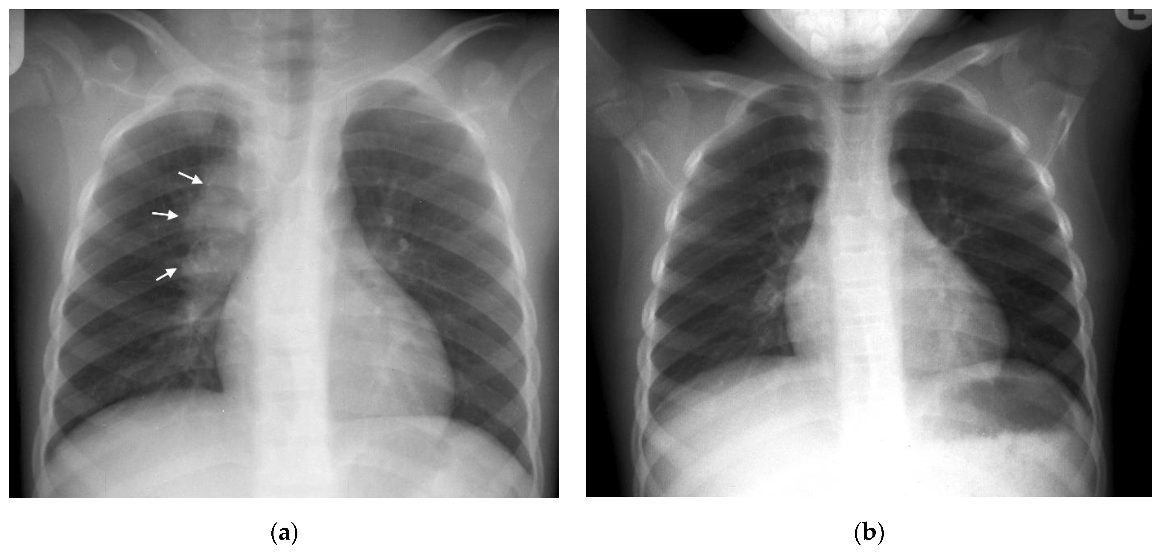
A Chest X Ray Of 3 Months Old Infant With Clinical Suspicion Of Mild Download Scientific Diagram

A Pediatric Chest X Ray With The Pb Shield The Circles Are The Download Scientific Diagram

Normal Pediatric Chest Xray Stock Photo Download Image Now Istock

What Is An X Ray For Kids Parents If Your Child Is Having An X Ray Exam This Is The Information They And You Too Will N Radiology X Ray Medical Imaging

Mengenal Cara Kerja Pemeriksaan X Ray Dan Efek Sampingnya Alodokter

A Chest X Ray Of 3 Months Old Infant With Clinical Suspicion Of Mild Download Scientific Diagram

A Pediatric Chest X Ray With The Pb Shield The Circles Are The Download Scientific Diagram

Medical Photography Radiology Humor Medical History

Chest X Ray Spot Diagnosis Chart Abnormal Heart Very Important For Health Professional Working In Radiolog Medical Anatomy Medical Knowledge Emergency Nursing